


PROTISTS
Protist Diversity
Before beginning this lab activity, be sure that you have read through the lab handout.
Now, using the images and videos below, you will sketch 9 different protists, each belonging to one of the major groups discussed in class. You can print and draw on the lab handout itself OR you may do so on a blank sheet of paper. If you have a tablet that you take notes/draw on then you may also draw digitally.
It is okay if you aren't an artist- after all this is biology lab! Just take your time and try to draw the 1) shape of the organism as best you can and 2) draw and label any distinctive features, such as cilia, flagella, etc. Focus on those features that are discussed in the lab handout. You do not have to color your drawings- but you can if you want.
Use the voicethread and/or the lab handout to list 1-2 characteristics for each organism. List these next to your drawing. When you are done, take a photo of your handout and upload to this week's discussion board along with the answers to the discussion questions. Contact your lab instructor with any questions.
Group: Protozoans
Sub-group: Flagellates
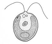
Organism: Giardia
Label: Flagella, nuclei


Look at the labeled diagram above. While the two large structures near the top of Giardia may look like eyeballs- they aren't! They are actually two nuclei that house the single-celled organism's genetic material.
Referring to the lab handout, what structure(s) does Giardia use to swim or move around its habitat?
Be sure to label these structures in your drawing.
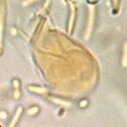
Group: Protozoans
Sub-group: Amoeba
Organism: Amoeba
Label: pseudopodia

Referring to the lab handout, what temporary structure does this organism use to move about its environment AND feed on prey?
Be sure to label this structure in your drawing.

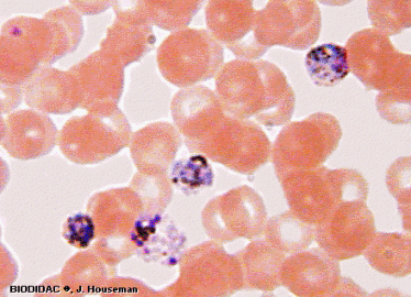
Group: Protozoans
Sub-group: Apicomplexans
Organism: Plasmodium
Label: Apex


Plasmodium infected cells (purple) among healthy red blood cells.
Close-up view of plasmodium (blue) using its apex to enter a host cell (red blood cell).
Virtually all apicomplexans are parasitic- meaning that they infect other host organisms and may cause disease. What structure do apicomplexans use to enter host cells and tissues?
Be sure to label this structure in your drawing.
Group: Protozoans
Sub-group: Ciliates
Organism: Paramecium
Label: cilia and oral groove


Referring to the lab handout, what structure(s) does this organism use to move about its environment AND feed on prey?
Be sure to label this structure(s) in your drawing.
Group: Slime Molds
Organism: Plasmodial Slime Molds
Label: plasmodium and fruiting body

To feed, the slime mold creates a plasmodium that seeks out food and water. The plasmodium and grow and recede as needed.
When conditions are tough (lack of food and water) then the plasmodium will develop into a fruiting body which will create and release spores. The spores will be spread around the environment and will lead to the next generation of plasmodial slime molds.
In your drawing, sketch out both parts of the life cycle of this organism, including the plasmodium and the fruiting body.

Group: Slime Molds
Organism: Cellular Slime Molds
Label: solitary cell, colony, and fruiting body
2) Solitary cells swarm to form a "slug"
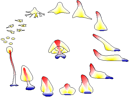
1) Solitary "amoeboa-like" cells
4) Spores develop into the next generation of solitary cells
3) The "slug" portion gives rise to a reproductive stalk, which will release reproductive spores into the environment.
Celllular slime molds behave differently compared to plasmodial slime molds.
Cellular slime molds go through a 'solitary phase' in which the single cells live independently. They will explore the environment and feed on their own.
When conditions get tough (lack of food and water) they solitary cells will swarm together to form a slug-like colony (shown on the right).
The colony can move together around the environment. Eventually, it will produce a fruiting body, which will produce spores. The spores will turn into the next generation of slime molds.
In your drawing, sketch out the major stages of the life cycle, including: the solitary cells-> the colony -> the fruiting body.

The slug-like form of the cellular slime mold.

Group: Algae
Organism: Dinoflagellates
Label: flagella


Red tide is a phenomenon caused by a red algae "bloom." Often, red tide is caused by various species of dinoflagellates. Red tide can decimate marine ecosystems by killing the marine life that lives there.
Label the flagella in your drawing.
Group: Algae
Organism: Diatoms

Diatoms have silica in their cell walls.
They have a glass-like appearance.
They come in a variety of shapes as shown to the right.


Group: Algae
Organism: Green Algae
Label: chloroplasts

This particular type of green algae is called 'spirogyra'. It is known for its spiral-shaped coils of chloroplasts (green).
Label the chloroplasts in your drawing.
